Respiratory muscles
Functional groups
The diagram below illustrates the respiratory muscles, grouped according to their roles in inspiration and forced expiration.
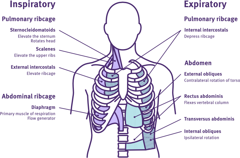
Respiratory muscles
Adapted from Patel et al. (2022).
Innervation and roles
The image and table below outline the respiratory muscles, their spinal nerve root innervations, and their roles in both ventilation and other functions.
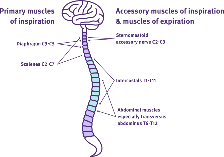
Respiratory muscles and spinal nerve root innervations
Adapted from SCIRE Professional
Muscle group:
- primary inspiratory muscle.
Innervation:
- phrenic nerve C3-C5.
Ventilation role:
- quiet and forced inspiration.
Other functions:
- generates dynamic transdiaphragmatic pressures by co-contract with the abdominal, internal intercostal, pelvic floor and intrinsic laryngeal muscles to regulate air flow for forced expiratory and body functions e.g. sighing, coughing, defecating
- aids venous and lymphatic return
- supports lower oesophageal sphincter function
- assists dynamic postural stability.
Muscles:
- internal intercostals
- external intercostals.
Muscle group:
- accessory inspiratory muscle
- expiratory muscle.
Innervation:
- spinal nerves T1-T11.
Ventilation role:
- quiet and forced inspiration (external intercostals only)
- forced expiration (internal intercostals only)
- forced expiratory actions e.g. cough.
Other functions:
- stabilises the rib cage and intercostal spaces when the diaphragm is contracting
- supports diaphragm function for efficient contraction.
Muscles:
- rectus abdominus
- external obliques
- internal obliques
- transversus abdominus.
Muscle group:
- accessory inspiratory muscle
- primary expiratory muscles.
Innervation:
- spinal nerves T6-T12.
Ventilation role:
- forced expiration
- forced expiratory actions e.g. cough.
Other functions:
- supports diaphragm function for efficient contraction—especially in sitting
- activates posterior pelvic tilt and trunk flexion movements
- facilitates postural stability for static and dynamic postural reactions.
Muscles and innervations:
- upper trapezius – accessory cranial nerve XI
- sternocleidomastoid – accessory cranial nerve XI and spinal nerve C2 and C3
- scalenes – spinal nerves C2-C7
- serratus anterior – long thoracic nerve of brachial plexus C5-C7
- pectoralis minor and major – lateral/medial pectoral nerves of brachial plexus C5-T1
- latissimus dorsi – thoracodorsal nerve C6-C8.
Other accessory muscles include the deep thoracic and paraspinal muscles.
Muscle group:
- accessory inspiratory muscles
- accessory expiratory muscles.
Ventilation role:
- forced inspiration
- forced expiration
- cough.
Other functions:
- activates head, neck, scapular and upper limb movements or stabilisation.
Physiopedia—muscles of respiration
physio-pedia.com
Respiratory management (rehab phase)
SCIRE Professional
Harvey, L. A. (2008). Management of spinal cord injuries: A guide for physiotherapists. Churchill Livingstone Elsevier.
Patel, N., Chong, K., & Baydur, A. (2022). Methods and applications in respiratory physiology: Respiratory mechanics, drive and muscle function in neuromuscular and chest wall disorders. Frontiers in Physiology, 13, Article 838414. https://doi.org/10.3389/fphys.2022.838414